Trends in Clinical and Medical Sciences
ISSN: 2791-0814 (online) 2791-0806 (Print)
DOI: 10.30538/psrp-tmcs2022.0026
The role of intra-abdominal pressure in injuries of the abdominal organs with associated injuries
Ishnazar Boynazarovich Mustafakulov\(^{1,*}\), Sobirjon Ergashevich Mamarajabov\(^{2}\) and Zilola Aramovna Djurayeva\(^{3}\)
\(^{1}\) Docent., Head of the Department of Surgical Diseases Samarkand, Uzbekistan.
\(^{2}\) Head of the Department of Operative Surgery and Topographic Anatomy of Samarkand State Medical University, Dean of the Faculty of International Education, Ph.D.; Samarkand, Uzbekistan.
\(^{3}\) Lecturer of the Department of Endocrinology, Samarkand, Uzbekistan.
Correspondence should be addressed to Ishnazar Boynazarovich Mustafakulov at med.author@yandex.ru
Abstract
Keywords:
1. Introduction
The main cause of death in closed abdominal trauma is the development of multiple organ failure [1,2,3,4,5,6]. Intra-abdominal hypertension (AHI) syndrome occupies an important place in the pathogenesis of multiple organ failure [7,8,9]. It is known that an increase in AHI contributes to an increase in the mortality rate, primarily in extremely severe patients [10]. The central link in the pathogenetic treatment of AHI syndrome is surgical aid. In connection with the above, this study analyzed the effect of various methods of surgical treatment on changes in the intra-abdominal pressure (IAP) in the postoperative period [11,12,13,14].
The ''gold standard'' for indirect measurement of intra-abdominal hypertension is the use of the bladder. Since the wall of the bladder is well stretchable and elastic with a volume of no more than 25 ml, it acts as a passive membrane and shows intra-abdominal pressure until it is true [15,16,17,18].
The purpose of the study was the timely prediction of the development of AHI syndrome in patients with closed abdominal trauma.
Material and research methods: All patients with closed abdominal trauma underwent measurement of intra-abdominal pressure and abdominal perfusion pressure (APP), for which a urethral catheter connected to a water pressure gauge of the Waldman apparatus was used. In this case, the IAP was examined every 6 hours. The APP was calculated by subtracting the IAP from the mean arterial pressure (MAP). SBP was determined using the following formula: SBP = (2 * diastolic blood pressure + systolic blood pressure) / 3.
The World Society developed the classification system for the Study of Abdominal Compartment Syndrome for AHI. Classification of AHI by degrees is as follows:
Grade I: IAD 12-15 mm Hg.;
II degree: IAD 16-20 mm Hg.;
III degree: IAD 21-25 mm Hg.;
IV degree: IAD> 25 mm Hg.
A normal APD is 60 mmHg. or higher.
2. Materials and methods
All patients were divided into the following groups depending on the method of completing the surgery. Group-I corresponded to the patients of the control group, which included 208 patients in whom the laparotomy was completed by traditional suturing of the wound tightly with drainage of the abdominal cavity. Group-II (main) was divided into two subgroups: the first subgroup (IIa) included 152 patients in whom laparotomy ended with the temporary closure of the surgical wound; the second subgroup (IIB) included 87 patients whose surgical wound was not sutured at the end of the operation, and a laparostomy was applied. All groups of patients under consideration were matched by sex, age, and severity of the condition according to the APACHE II scale and the scale for determining the severity of injury (ISS).3. Results and discussion
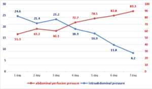
AHI of I degree was observed in 271 (56.7%) patients, II degree - in 167 (35.0%), III degree - in 28 (5.8%), and IV degree - in 12 (2.5%) patients. ... In patients with concomitant abdominal trauma, a decrease in APD was noted below 80 mm Hg. Art. in 70.3% of cases, while the APD level is less than 65 mm Hg. Art. was noted in 28.9% of cases. In all groups of victims with concomitant abdominal trauma in the postoperative period, we analyzed the results of studying the dynamics of APD and IAP.On the first day of the postoperative period, the patients of group I (traditional tactics, Figure 1) had a second-degree AHI with a critical increase in the level of intra-abdominal pressure by day 4 (20.9 \(\pm\) 3.8 mm Hg). At the same time, APD values remained low, reaching the minimum values (62.8 \(\pm\) 8.7 mm Hg) by day 3 of the postoperative period. In the dynamics against treatment background in the first week, in 58.7% of cases, there was a gradual decrease in IHD and normalization of APD indicators. In 94 (45.2%) patients, various complications developed (incompetence of the inner intestinal anastomosis, eventration of the abdominal organs, enclosed abscesses of the abdominal cavity), gastroduodenal bleeding, early adhesive obstruction), which were the reason for the reoperation.
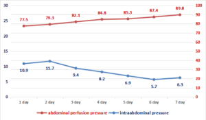
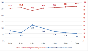
In subgroups IIa and IIb of groups (damage control surgery, Figures 2 and 3), a statistically significant decrease in IAP were observed on the initial day of the postoperative period: up to 10.9 \(\pm\) 7.8 mm Hg. Art. in subgroup IIa and up to 7.8 \(\pm\) 3.5 mm Hg. Art. in subgroup IIb (p> 0.05). There is an improvement in APD indicators - up to 77.5 \(\pm\) 5.8 mm Hg. Art. and 84.5 \(\pm\) 4.2 mm Hg. Art. respectively (p> 0.05), which shows an improvement in microcirculation processes in the main group's victims. Moreover, the increase in IAP in the IIa subgroup of patients on the third day of the postoperative period to 12.3 \(\pm\) 3.7 mm Hg. Art. should not be misleading since this depends on the second stage of surgical tactics and the final closure of the surgical wound.
Figure 1. Changes in indicators of abdominal perfusion pressure and intra-abdominal pressure in patients of the control group (early total care).
Figure 2. Changes in indicators of abdominal perfusion pressure and intra-abdominal pressure in patients of the main group (damage control surgery) with temporary closure of the anterior abdominal wall.
Figure 3. Changes in indicators of abdominal perfusion pressure and intra-abdominal pressure in patients of the main group (damage control surgery) with the imposition of a laparostomy.
The frequency of the development of multiple organ failure according to the criteria and the assessment on the SOFA scale were compared on the second day of the postoperative period since it was during this period that the most significant difference in IAP indicators was revealed between I and IIa, and I and IIb groups (Table 1).
Table 1. Multiple organ failure and SOFA score in victims on the 2nd day of the postoperative period.
| Groups Systems | Group I, n = 208 | Subgroup IIa, n = 152 | Subgroup IIb, n = 87 |
|---|---|---|---|
| The cardiovascular system | 22.5\% * | 55.6% | 33.3% |
| urinary system | 45% * | 22.2% | 22.2% |
| Respiratory system | 85% | one hundre | 63.0% |
| Liver |
- |
- | |
| Coagulating system | 10% | 22.2% | 3.7% |
| Metabolic dysfunction | 32.5% * | 11.1% | 7.40% |
| CNS | 87.5% | 77.8% | 17.4% |
| SOFA, score | 7.3 \(\pm\) 1.8 * | 3.4 \(\pm\) 1.5 | 3.7 \(\pm\) 1.8 |
The high incidence of acute cerebral failure in all groups is explained by the nature of the trauma, that is, abdominal trauma was almost always combined with craniocerebral trauma, which was the cause of neurological deficit on the Glasgow coma scale. Manifestations of hepatic impairments were not observed in any of the cases, and impairments of the coagulating system were also rare and did not differ significantly among the groups. But nevertheless, in the first group there were significantly more patients with disorders in the cardiovascular, urinary, and metabolic disorders. This group of patients really needed more inotropic therapy. The result of the SOFA assessment showed a truly higher score in the severity of organ disorders in the first group.
The overall mortality rate in patients with abdominal injuries and associated severe trauma was 67.8%. The number of deaths among the patients of the first group was 119 out of 208 (57.21%), in the second group - 88 out of 270 (32.59%). Differences between IIa and IIb subgroups are not significant (p < 0.05). However, the differences in indicators I and IIa, I and IIb by subgroups are significant (\(p < 0.05\)).
Treatment results were studied in absolutely all patients with different IAP parameters during admission to the ICU. The average IAP in those patients who survived was 8.5 \(\pm\) 3.2 mm Hg, and in deceased patients - 24.2 \(\pm\) 1.8 mm Hg. (p < 0.05). But in this case, a natural dynamics of an increase in the mortality rate with an increase in IAP was noted.
When analyzing the relationship between AHI, APD, the frequency of multiple organ failure and the general severity of the condition of the victims, a statistically significant correlation was confirmed between the IAP and the severity of the patient's condition according to the SOFA and APACHE II scales (\(p < 0.05\)). The increase in IAP level corresponded to the worsening of the severity of the condition of the victims according to the SOFA and APACHE II scales, which was confirmed by the progress of the clinical picture of multiple organ failure.
4. Conclusion
Thus, in order to timely predict the development of AHI syndrome in patients with closed abdominal trauma, it is necessary to monitor the IAP level. IBH syndrome develops in patients with concomitant abdominal trauma and is characterized by relatively high mortality rates. A statistically significant correlation was established between the level of AHI, APD, the frequency of development of a picture of multiple organ failure and the severity of the patient's condition according to the SOFA and APACHE II scales (p < 0.05). A sudden increase and persistence of a high IAP level for a long time in patients with a closed abdominal trauma is an indication for the use of active surgical tactics to perform decompression.Author Contributions:
All authors contributed equally to the writing of this paper. All authors read and approved the final manuscript.Conflicts of Interest:
''The authors declare no conflicts of interest.''References
- Shakirov, B. M. (2022). The role of intra-abdominal pressure in injuries of the ab-dominal organs with associated injuries. International Journal of Surgery and Transplantation Research, 2(1), 1-5.[Google Scholor]
- Altyev, B. K., & Rakhimov, O. U. (2018). Intraabdominal bleedings after various options of cholecystectomy. Central Asian Journal of Medicine, 2018(1), 14-19. [Google Scholor]
- Valiev, E. Y. (2011). Experience in providing specialized care to patients with polytrauma in the conditions of the RSCEMP. collection. ''Modern military field surgery and surgery of injuries.'' St. Petersburg, 67-68. [Google Scholor]
- Coccolini, F., Kobayashi, L., Kluger, Y., Moore, E. E., Ansaloni, L., Biffl, W., ... & Coimbra, R. (2019). Duodeno-pancreatic and extrahepatic biliary tree trauma: WSES-AAST guidelines. World Journal of Emergency Surgery, 14, Article No. 56. https://doi.org/10.1186/s13017-019-0278-6. [Google Scholor]
- Pereira, R., Vo, T., & Slater, K. (2019). Extrahepatic bile duct injury in blunt trauma: A systematic review. Journal of Trauma and Acute Care Surgery, 86(5), 896-901. [Google Scholor]
- Ikramov, A. I., & Khalibaeva, G. B. (2019). Radiology diagnostics of bladder and urethral injuries in pelvic trauma. Medical Visualization, 16(2), 109-118. [Google Scholor]
- Gafurovich, N. F., Babajanovich, K. Z., Mirolimovich, A. M., & Azadovich, A. P. (2018). Results of surgical treatment of ''fresh'' injuries of magistral bile ducts. European Science Review, 2018(7-8), 148-152. [Google Scholor]
- Mustafakulov, I. B., Umedov, K. A., Karabaev, H. K., & Djuraeva, Z. A. (2021). Damage Control the Liver and Spleen in Case of Concomitant Injury (Literature Review). Advances in Clinical Medical Research, 2(2), 13-17.[Google Scholor]
- Gavrishuk, Y. V., Kazhanov, I. V., Tulupov, A. N., Demko, A. E., Kandyba, D. V., Mikityuk, S. I., & Kolchanov, E. A. (2019). Minimally invasive treatment in the victim with spleen injury. Grekov's Bulletin of Surgery, 178(4), 58-60. [Google Scholor]
- Mohan, B., Bhoday, H. S., Aslam, N., Kaur, H., Chhabra, S., Sood, N., & Wander, G. (2013). Hepatic vascular injury: clinical profile, endovascular management and outcomes. Indian Heart Journal, 65(1), 59-65. [Google Scholor]
- Mustafakulov, I. B., & Djuraeva, Z. A. (2019). Intra-abdominal hypertension at combined injuries of the abdominal organs. American Journal of Medicine and Medical Sciences, 9(12), 499-502. [Google Scholor]
- Mustafakulov, I. B., & Djuraeva, Z. A. (2020). Severe associated trauma to the abdomen diagnosis and treatment. European Journal of Pharmaceutical and Medical Research, 7(6), 113-116. [Google Scholor]
- Mustafakulov, I. B., & Djuraeva, Z. A. (2020). Evaluaton of the effectiveness of multi-stage surgical tactics for liver damage. World Journal of Advance Healthcare Research, 4(3), 264-266. [Google Scholor]
- Mustafakulov, I. B., Elmuradov, A., Djuraeva, Z. A., & Umedov, H. A. (2021). Diagnostic tools and therapeutic possibilities of endovideolaparoscopy for combined abdominal trauma. Journal of Natural Remedies, 22(1(2)), 181-186. [Google Scholor]
- Ozogul, B., Kisaoglu, A., Aydinli, B., Öztürk, G., Bayramoglu, A., Saritemur, M., ... & Atamanalp, S. (2014). Non-operative management (NOM) of blunt hepatic trauma: 80 cases. Ulusal Travma Ve Acil Cerrahi Dergisi-Turkish Journal Of Trauma & Emergency Surgery, 20(2), 97-100.[Google Scholor]
- Saaiq, M., Niaz-ud-Din, Z. M., & Shah, S. A. (2013). Presentation and outcome of surgically managed lives trauma: experience at a tertiary care teaching hospital. Journal Of Pakistan Medical Association, 63(4), 436-9. [Google Scholor]
- Swaid, F., Peleg, K., Alfici, R., Olsha, O., Jeroukhimov, I., Givon, A., & Kessel, B. (2015). The severity of liver injury following blunt trauma does not correlate with the number of fractured ribs: an analysis of a national trauma registry database. Surgery today, 45(7), 846-850. [Google Scholor]
- Wang, Y. C., Fu, C. Y., Chen, Y. F., Hsieh, C. H., Wu, S. C., & Yeh, C. C. (2011). Role of arterial embolization on blunt hepatic trauma patients with type I contrast extravasation. The American Journal of Emergency Medicine, 29(9), 1147-1151. [Google Scholor]


